
 A study from the University of Eastern Finland and Massachusetts Institute of Technology hows that articular cartilage degenerates specifically around injury areas when the fluid flow velocity becomes excessive.
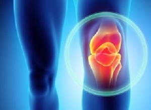
Knee joint injuries are typically related to sports, such as football, rugby or ice hockey, but people often do not know that such injuries may lead to joint inflammation and post-traumatic osteoarthritis. In advanced post-traumatic osteoarthritis, joint cartilage breaks down completely, causing severe joint pain, lack of mobility and even social isolation.
However, the mechanisms leading to osteoarthritis are not known. Currently, it is not possible for a physician examining a patient to predict future joint condition and possible development of osteoarthritis. In the future, however, this may be possible, as a study from the University of Eastern Finland and Massachusetts Institute of Technology now shows that articular cartilage degenerates specifically around injury areas when the fluid flow velocity becomes excessive.
The study presents a new mechano-biological model for cartilage degeneration by implementing tissue deformation and fluid flow as mechanisms for cartilage breakdown when a normal dynamic loading, such as walking, is applied to the joint. The results were compared to experimentally observed degradation of articular cartilage. Ultimately, the new model could be used to predict osteoarthritis in personal medicine, to suggest optimal rehabilitation protocols, and to improve the quality of life.
The researchers found that different mechanisms, such as fluid flow and tissue deformation, can cause cartilage degradation after a knee injury. The results obtained using the novel algorithm agreed well with the experimentally observed proteo-glycan content and cell death in cartilage samples. According to the researchers, a numerical analysis shows that both fluid flow and tissue deformation are plausible mechanisms leading to osteoarthritis, but increased fluid flow from cartilage seems to be better in line with the experiments.
“Our findings indicate that after an injury in the knee and subsequent tissue loading, osteoarthritis is caused by easy leakage of proteoglycans through the injury surface by high fluid outflow,” early stage researcher Gustavo A Orozco from the University of Eastern Finland explains.
The findings are significant, and could open up new avenues for the model to be employed in the prediction of subject-specific progression of post-traumatic osteoarthritis, and in the evaluation of the effect of clinical interventions in the future. Specifically, the model could identify high and low-risk lesions in the cartilage for osteoarthritis development and suggest an optimal and individual rehabilitation protocol.
The study has received funding from the EU’s Horizon 2020 research and innovation programme under the Marie Sklodowska-Curie grant No 713645.
Abstract
Cartilage provides low-friction properties and plays an essential role in diarthrodial joints. A hydrated ground substance composed mainly of proteoglycans (PGs) and a fibrillar collagen network are the main constituents of cartilage. Unfortunately, traumatic joint loading can destroy this complex structure and produce lesions in tissue, leading later to changes in tissue composition and, ultimately, to post-traumatic osteoarthritis (PTOA). Consequently, the fixed charge density (FCD) of PGs may decrease near the lesion. However, the underlying mechanisms leading to these tissue changes are unknown. Here, knee cartilage disks from bovine calves were injuriously compressed, followed by a physiologically relevant dynamic compression for twelve days. FCD content at different follow-up time points was assessed using digital densitometry. A novel cartilage degeneration model was developed by implementing deviatoric and maximum shear strain, as well as fluid velocity controlled algorithms to simulate the FCD loss as a function of time. Predicted loss of FCD was quite uniform around the cartilage lesions when the degeneration algorithm was driven by the fluid velocity, while the deviatoric and shear strain driven mechanisms exhibited slightly discontinuous FCD loss around cracks. Our degeneration algorithm predictions fitted well with the FCD content measured from the experiments. The developed model could subsequently be applied for prediction of FCD depletion around different cartilage lesions and for suggesting optimal rehabilitation protocols.
Authors
Gustavo A Orozco, Petri Tanska, Cristina Florea, Alan J Grodzinsky, Rami K Korhonen
[link url="https://www.nature.com/articles/s41598-018-33759-3"]Scientific Reports abstract[/link]