
 Researchers have developed a device using light scattering spectroscopy that in a study distinguished between harmless pancreatic cysts and those having malignant potential with an overall accuracy of 95%.
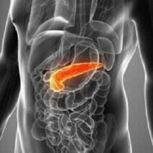
Pancreatic cancer has the lowest survival rate among all major cancers, largely because physicians lack diagnostic tools to detect the disease in its early, treatable stages. Now, a team of investigators led by Dr Lev T Perelman, director of the Centre for Advanced Biomedical Imaging and Photonics at Beth Israel Deaconess Medical Centre (BIDMC), has developed a promising new tool capable of distinguishing between harmless pancreatic cysts and those with malignant potential with an overall accuracy of 95%.
The new device uses light scattering spectroscopy (LSS) to detect the structural changes that occur in cancerous or pre-cancerous cells by bouncing light off tissues and analysing the reflected spectrum. The results could help guide physicians' decision making when considering whether the presence of pancreatic cysts requires surgery, a high-risk procedure. Today, because of the lack of less-invasive diagnostic methods, more than half of these procedures turn out to have been unnecessary.
"About one-fifth of pancreatic cancers develop from cysts, but not all lesions are cancerous," said Perelman, who is also professor of medicine and professor of obstetrics, gynaecology and reproductive biology at Harvard Medical School. "Considering the high risk of pancreatic surgeries and the even higher mortality from untreated pancreatic cancers, there's an obvious need for new diagnostic methods to accurately identify the pancreatic cysts that need surgical intervention and those that do not."
In Perelman and colleagues' series of experiments, the LSS technique achieved 95% accuracy for identifying malignancy. Cytology – the only pre-operative test currently availably – is accurate only 58% of the time. While the new technique requires further testing, LSS could represent a major advance against pancreatic cancer.
"This tool is a technology that is transformative in the evaluation of pancreatic cysts," said co-lead author Dr Douglas K Pleskow, clinical chief of the division of gastroenterology and director of the colon and rectal cancer programme at the Cancer Centre at BIDMC. "It provides a high level of precision in the detection of potential malignant transformation of these cysts."
Pancreatic cysts are common, and today's high-definition scanning technologies like MRI and CT imaging are detecting them with increasing frequency. Despite their high resolution, these scanners provide doctors with limited information about cysts' malignant potential.
Currently, physicians rely on minimally-invasive fine needle aspiration (FNA) biopsies to test pancreatic cysts for malignancy. The biopsy removes fluid from the cysts, which is then analyzed for cancer cells and other telltale signs of the disease, a process called cytology. However, the test fails to detect cancer about half the time, leaving high-risk surgery as the current gold-standard means of diagnosing pancreatic cysts.
To test the accuracy of the LSS system, Perelman and colleagues collected and analysed the reflected light from 13 cysts taken from recent surgeries. Next, they compared their findings with the results from pre-operative imaging, FNA biopsies and post-operative tissues analysis. In all cases, the LSS diagnosis agreed with the post-operative analysis.
In a second experiment, the LSS tool was tested in 14 patients with pancreatic cysts who were undergoing the standard FNA biopsy. Measuring less than half a millimeter in diameter, the miniature experimental LSS fibre-optic probe was inserted in the FNA needle. Physicians spent two minutes or less measuring optical spectra from the internal cyst surface before collecting fluid from the cysts as part of the traditional biopsy. Out of nine patients whose cysts had been definitely diagnosed as either cancerous or benign, all were correctly identified by LSS.
Next, the researchers will assess the LSS system's accuracy by continuing to analyse post-operative tissues as they become available.
Abstract
Pancreatic cancers are usually detected at an advanced stage and have poor prognosis. About one-fifth of these arise from pancreatic cystic lesions. Yet not all lesions are precancerous, and imaging tools lack adequate accuracy for distinguishing precancerous from benign cysts. Therefore, decisions on surgical resection usually rely on endoscopic ultrasound-guided fine-needle aspiration (EUS-FNA). Unfortunately, cyst fluid often contains few cells, and fluid chemical analysis lacks accuracy—which has dire consequences, including unnecessary pancreatic surgery for benign cysts and the development of cancer. Here, we report an optical spectroscopic technique, based on a spatial gating fibre-optic probe, that predicts the malignant potential of pancreatic cystic lesions during routine diagnostic EUS-FNA procedures. In a double-blind prospective study in 25 patients, with 14 cysts measured in vivo and 13 postoperatively, the technique achieved an overall accuracy of 95%, with a 95% confidence interval of 78–99%, in cysts with definitive diagnosis.
Authors
Lei Zhang, Douglas K Pleskow, Vladimir Turzhitsky, Eric U Yee, Tyler M Berzin, Mandeep Sawhney, Shweta Shinagare, Edward Vitkin, Yuri Zakharov, Umar Khan, Fen Wang, Jeffrey D Goldsmith, Saveli Goldberg, Ram Chuttani, Irving Itzkan, Le Qiu, Lev T Perelman
[link url="http://www.bidmc.org/News/PRLandingPage/2017/March/Perelman-Pleskow.aspx"]Beth Israel Deaconess Medical Centre material[/link]
[link url="http://www.nature.com/articles/s41551-017-0040"]Nature Biomedical Engineering abstract[/link]