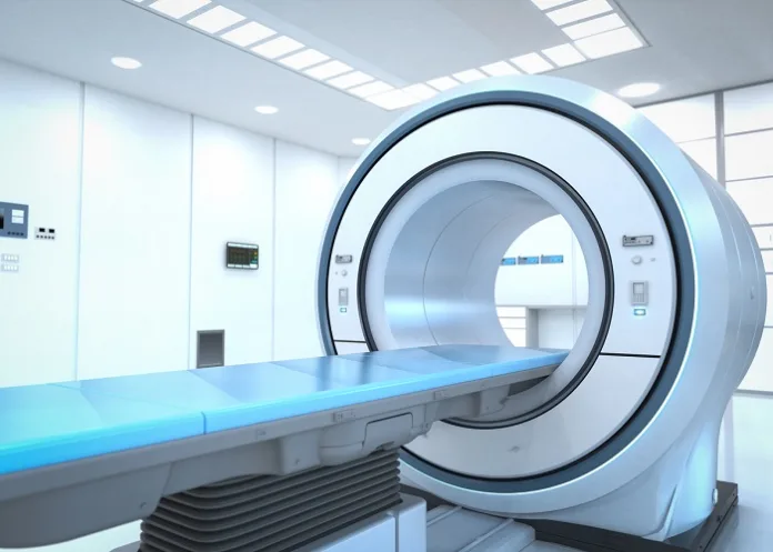
Perseverance and ingenuity have paid off for Hong Kong researchers, who, like numerous others, have been exploring options for a low-field MRI system with a magnetic field strength of less than 1 T as an alternative to the loud, expensive machines requiring special rooms with shielding to block their powerful magnetic field.
Most low-field scanners in development are for brain scans only. In 2022, the US Food and Drug Administration (FDA) cleared the first portable MRI system: Hyperfine’s Swoop, designed for use at a patient’s bedside – for head and brain scans.
But the technology has not been applied to whole-body MRI, until now.
This month, scientists from The University of Hong Kong published a study in Science describing a whole-body, ultra low–field MRI, which experts have described as a breakthrough.
The device uses a 0.05 T magnet – one 60th the magnetic field strength of the standard 3 T MRI model common in hospitals today, said lead author Ed Wu, PhD, professor of biomedical engineering at The University of Hong Kong.
Medscape reports that because the field strength is so low, no protective shielding is needed. Patients and bystanders can safely use smartphones. And the scanner is safe for patients with implanted devices, like a cochlear implant or pacemaker, or any metal on their body or clothes. No hearing protection is required, either, because the machine is so quiet.
If all goes well, the technology could be commercially available in as little as a few years, Wu said.
But first, funding and FDA approval would be needed.
“A company is going to have to come along and say, ‘This looks fantastic. We’re going to commercialise this and go through this certification process’,” said Andrew Webb, PhD, professor of radiology and the founding director of the Gorter MRI Centre at the Leiden University Medical Centre, Netherlands. (Webb was not involved in the study.)
Kevin Sheth, MD, director of the Yale Centre for Brain & Mind Health, who was also not involved in the study, said: “This is a breakthrough. It is one of the first, if not the first, demonstrations of low-field MRI imaging for the entire body.”
Improving access to MRI
One hope for this technology is to bring MRI to more people worldwide. Africa has less than one MRI scanner per million residents, whereas the United States has about 40.
While a new 3 T machine can cost about $1m, the low-field version is much cheaper – only about $22 000 in materials cost per scanner, according to Wu.
A low magnetic field means less electricity, too: the machine can be plugged into a standard wall outlet. And because a fully shielded room isn't needed, that could save another $100 000 in materials, Webb said, adding that its ease of use could improve accessibility in countries with limited training.
“To be a technician is two to three years of training for a regular MRI machine, a lot of it to do with safety, a lot of it to do very subtle planning. These (low-field) systems are much simpler.”
Challenges and the future
The prototype weighs about 1.5 tons (a 3 T MRI can weigh between 6 and 13 tons). That might sound like a lot, but it’s comparable to a mobile CT scanner, which is designed to be moved from room to room.
“Plus, its weight can be substantially reduced if further optimised,” Wu said.
One challenge with low-field MRIs is image quality, which tends to be not as clear and detailed as those from high-power machines. To address this, the research team used deep learning (artificial intelligence) to enhance the image quality.
“Computing power and large-scale data underpin our success, which tackles the physics and math problems that are traditionally considered intractable in existing MRI methodology.”
Webb said he was impressed by the image quality shown in the study. “They look much higher quality than you would expect from such a low-field system,” he said.
Still, only healthy volunteers were scanned. The true test will be using it to view subtle pathologies, he added.
That’s what Wu and his team are working on now – taking scans to diagnose various medical conditions. His group’s brain-only version of the low-field MRI has been used for diagnosis, he noted.
Study details
Whole-body magnetic resonance imaging at 0.05 Tesla
Yujiao Zhao, Ye Ding, Vick Lau, Christopher Man, Ed X. Wu et al.
Published in Science on 10 May 2024
Abstract
Introduction
Magnetic resonance imaging (MRI) has revolutionised healthcare with its nonionising, non-invasive, multicontrast, and quantitative capabilities. It also presents a promising platform for future artificial intelligence–driven medical diagnoses. However, after five decades of development, MRI accessibility, especially in low and middle-income countries, remains low and highly uneven due to high costs and specialised settings required for standard superconducting MRI scanners. These scanners are mostly found in specialised radiology departments and large imaging centres, restricting their availability in other medical settings. The need for radio frequency (RF)-shielded rooms and high power consumption further adds to hardware cost and compromises mobility and patient-friendliness.
Rationale
We developed a highly simplified whole-body ultra-low-field (ULF) MRI scanner that operates on a standard wall power outlet without RF or magnetic shielding cages. This scanner uses a compact 0.05 Tesla permanent magnet and incorporates active sensing and deep learning to address electromagnetic interference (EMI) signals. We deployed EMI sensing coils positioned around the scanner and implemented a deep learning method to directly predict EMI-free nuclear magnetic resonance signals from acquired data. To enhance image quality and reduce scan time, we also developed a data-driven deep learning image formation method, which integrates image reconstruction and three-dimensional (3D) multiscale super-resolution and leverages the homogeneous human anatomy and image contrasts available in large-scale, high-field, high-resolution MRI data.
Results
We implemented commonly used clinical protocols at 0.05 Tesla, including T1-weighted, T2-weighted, and diffusion-weighted imaging, and optimised their contrasts for different anatomical structures. Each protocol was designed to have a scan time of eight minutes or less with an image resolution of approximately 2×2×8 mm³. The scanner power consumption during scanning was under 1800W and around 300W when idle. We conducted imaging on healthy volunteers, capturing brain, spine, abdomen, lung, musculoskeletal, and cardiac images. Deep learning signal prediction effectively eliminated EMI signals, enabling clear imaging without shielding. The brain images showed various brain tissues whereas the spine images revealed intervertebral disks, spinal cord, and cerebrospinal fluid. Abdominal images displayed major structures like the liver, kidneys, and spleen. Lung images showed pulmonary vessels and parenchyma. Knee images identified knee structures such as cartilage and meniscus. Cardiac cine images depicted the left ventricle contraction and neck angiography revealed carotid arteries. Furthermore, deep learning image formation greatly improved the 0.05 Tesla image quality for various anatomical structures, including the brain, spine, abdomen, and knee; it also effectively suppressed noise and artefacts and increased image spatial resolution.
Conclusion
To address MRI accessibility challenges, we developed a low-power and simplified whole-body 0.05 Tesla MRI scanner that operates without the need for RF or magnetic shielding and that can be manufactured, maintained, and operated at a low cost. We experimentally demonstrated the general utility of this scanner for imaging various human anatomical structures at a whole-body level, even in the presence of strong EMI signals, with acceptable scan time. Moreover, we demonstrated the potential of deep learning image formation to substantially augment 0.05 Tesla image quality by exploiting computing and extensive high-field MRI data. These advances pave the way for affordable, patient-centric, and deep learning–powered ULF MRI scanners, addressing unmet clinical needs in diverse healthcare settings worldwide
Science article – Whole-body magnetic resonance imaging at 0.05 Tesla (Open access)
Medscape article – 'Big Breakthrough': New Low-Field MRI Is Safer and Easier (Open access)
See more from MedicalBrief archives:
Portable MRIs almost as effective as standard MRIs in detecting strokes
Hand-held ultrasound scanner shows its potential in rural Africa
Mpumalanga patient waits more than a year for MRI

