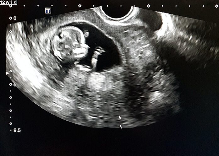
A routine prenatal ultrasound in the second trimester can identify early signs of Autism Spectrum Disorder (ASD), a study by Ben-Gurion University of the Negev and Soroka Medical Center found.
The researchers examined data from hundreds of prenatal ultrasound scans from the foetal anatomy survey conducted during mid-gestation. They found anomalies in the heart, kidneys, and head in 30% of foetuses who later developed ASD, a three-times higher rate than was found in typically developing foetuses from the general population. and twice as high as their typically developing siblings.
Anomalies were detected more often in girls than in boys and the severity of the anomalies was also linked to the subsequent severity of ASD.
Researchers from the Azrieli National Centre for Autism and Neurodevelopment Research published their findings in the peer-reviewed journal Brain. Prof Idan Menashe, a member of the Centre and the Department of Public Health in the Faculty of Health Sciences, led the research with his MD/PhD student Ohad Regev.
“Doctors can use these signs, discernible during a routine ultrasound, to evaluate the probability of the child being born with ASD,” said Menashe. “Previous studies showed that children born with congenital diseases, primarily those involving the heart and kidneys, had a higher chance of developing ASD. Our findings suggest that certain types of ASD that involve other organ anomalies, begin and can be detected, in utero.”
A previous study by the Centre found early diagnosis and treatment increased social ability by three times as much. Prenatal diagnosis could mean a course of treatment from birth instead of waiting until age two or three or even later.
Study details
Association between ultrasonography foetal anomalies and autism spectrum disorder
Ohad Regev, Amnon Hadar, Gal Meiri, Hagit Flusser, Analya Michaelovski, Ilan Dinstein, Reli Hershkovitz, Idan Menashe.
Published in Brain on 17 January 2022
Abstract
Multiple evidence support the prenatal predisposition of autism spectrum disorder (ASD). Nevertheless, robust data about abnormalities in foetuses later developing into children diagnosed with ASD are lacking. Prenatal ultrasound is an excellent tool to study abnormal fetal development as it frequently used to monitor foetal growth and identify foetal anomalies throughout pregnancy.
We conducted a retrospective case-sibling-control study of children diagnosed with ASD (cases); their own typically developing, closest-in-age siblings (TDS); and typically developing children from the general population (TDP), matched by year of birth, sex and ethnicity to investigate the association between ultrasonography foetal anomalies (UFAs) and ASD. The case group was drawn from all children diagnosed with ASD enrolled at the Azrieli National Center of Autism and Neurodevelopment Research. Foetal ultrasound data from the foetal anatomy survey were obtained from prenatal ultrasound clinics of Clalit Health Services (CHS) in southern Israel.
The study comprised 659 children: 229 ASD, 201 TDS, and 229 TDP. UFAs were found in 29.3% of ASD cases vs. only 15.9% and 9.6% in the TDS and TDP groups (aOR = 2.23, 95%CI = 1.32-3.78, and aOR = 3.50, 95%CI = 2.07-5.91, respectively). Multiple co-occurring UFAs were significantly more prevalent among ASD cases. UFAs in the urinary system, heart, and head & brain were the most significantly associated with ASD diagnosis (aORUrinary =2.08, 95%CI = 0.96-4.50 and aORUrinary = 2.90, 95%CI = 1.41-5.95; aORHeart = 3.72, 95%CI = 1.50-9.24 and aORHeart = 8.67, 95%CI = 2.62-28.63; and aORHead&Brain = 1.96, 95%CI = 0.72-5.30 and aORHead&Brain = 4.67, 95%CI = 1.34-16.24; vs. TDS and TDP, respectively). ASD females had significantly more UFAs than ASD males (43.1% vs. 25.3%, p = 0.013) and a higher prevalence of multiple co-occurring UFAs (15.7% vs. 4.5%, p = 0.011). No sex differences were seen among TDS and TDP controls. ASD foetuses were characterised by a narrower head and a relatively wider ocular-distance vs. TDP foetuses (ORBPD = 0.81, 95%CI = 0.70-0.94, and aOROcular-Distance = 1.29, 95%CI = 1.06-1.57). UFAs were associated with more severe ASD symptoms.
Our findings shed important light on the abnormal multiorgan embryonic development of ASD and suggest foetal ultrasonography biomarkers for ASD.
See more from MedicalBrief archives:
Landmark intervention may reduce autism diagnosis rates by two-thirds
AI and machine learning to detect early autism through retina scans
Autism so over-diagnosed that term is becoming meaningless — study
EEGs accurately diagnose autism in infants

