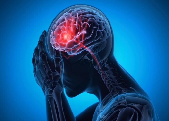
British researchers have suggested that severe Covid infections may drive inflammation in the brain’s “control centre”, causing damage that could explain the long-term breathlessness, fatigue and anxiety experienced by some patients.
High-resolution MRI scans of 30 people hospitalised with Covid early in the pandemic, before the introduction of vaccines, found signs of inflammation in the brainstem, a small but critical structure that governs life-sustaining bodily functions such as breathing, heart rate and blood pressure, they said.
The scans suggest that severe Covid infections can provoke an immune reaction which inflames the brainstem, with the resulting damage producing symptoms that can last for months after patients have been discharged.
“The fact that we see abnormalities in the parts of the brain associated with breathing strongly suggests that long-lasting symptoms are an effect of inflammation in the brainstem after Covid-19 infection,” said Dr Catarina Rua, a neuroscientist at the University of Cambridge and first author on the study.
The Guardian reports that the project was launched before researchers and public health officials knew about long Covid. But many people with long Covid report breathlessness and fatigue, raising the possibility that brain inflammation could be involved in their symptoms, too.
“We didn’t study people with long Covid, but they do often have long-lasting effects of breathlessness and fatigue, which are similar to the symptoms these very severely affected people had six months after they were hospitalised,” Rua said. “It does lead us to ask the question, do people with long Covid have any brainstem changes?”
Rua and her colleagues used powerful 7 Tesla MRI scanners to image the patients’ brains. These revealed enough detail to see inflammation and microstructural abnormalities in the brainstem tissue. All of the patients had been admitted to hospital with severe Covid near the start of the pandemic.
The scans highlighted abnormalities linked to inflammation in multiple parts of the brainstem, starting several weeks after patients were admitted to hospital. The damage was still evident in scans more than six months later, they wrote in the journal Brain.
Damage to the brainstem might also contribute to the mental health problems some patients face after Covid infection. Of the patients in the study, those with the highest levels of brainstem inflammation had the most severe physical symptoms and the highest levels of depression and anxiety, according to the study published in Brain.
“While this study does not conclusively prove the causes of long Covid, it does point a finger at one possible suspect for some of the symptoms experienced,” said Paul Mullins, a professor in neuroimaging at the University of Bangor. “It is not clear that this shows much in the way of possible treatments for long Covid once it has occurred, but it perhaps does point to the need to reduce inflammatory responses during initial Covid infection and response.”
Study details
Quantitative susceptibility mapping at 7 T in Covid-19: brainstem effects and outcome associations
Catarina Rua, Betty Raman, Christopher Rodgers et al.
Published in Brain on 7 October 2024
Abstract
Post-mortem studies have shown that patients dying from severe acute respiratory syndrome coronavirus (SARS-CoV-2) infection frequently have pathological changes in their CNS, particularly in the brainstem. Many of these changes are proposed to result from para-infectious and/or post-infection immune responses. Clinical symptoms such as fatigue, breathlessness, and chest pain are frequently reported in post-hospitalised coronavirus disease 2019 (Covid-19) patients. We propose that these symptoms are in part due to damage to key neuromodulatory brainstem nuclei. While brainstem involvement has been demonstrated in the acute phase of the illness, the evidence of long-term brainstem change on MRI is inconclusive. We therefore used ultra-high field (7 T) quantitative susceptibility mapping (QSM) to test the hypothesis that brainstem abnormalities persist in post-Covid patients and that these are associated with persistence of key symptoms.
We used 7 T QSM data from 30 patients, scanned 93–548 days after hospital admission for Covid-19 and compared them with 51 age-matched controls without prior history of Covid-19 infection. We correlated the patients’ QSM signals with disease severity (duration of hospital admission and COVID-19 severity scale), inflammatory response during the acute illness (C-reactive protein, D-dimer and platelet levels), functional recovery (modified Rankin scale), depression (Patient Health Questionnaire-9) and anxiety (Generalised Anxiety Disorder-7).
In Covid-19 survivors, the MR susceptibility increased in the medulla, pons and midbrain regions of the brainstem. Specifically, there was increased susceptibility in the inferior medullary reticular formation and the raphe pallidus and obscurus. In these regions, patients with higher tissue susceptibility had worse acute disease severity, higher acute inflammatory markers, and significantly worse functional recovery.
This study contributes to understanding the long-term effects of Covid-19 and recovery. Using non-invasive ultra-high field 7 T MRI, we show evidence of brainstem pathophysiological changes associated with inflammatory processes in post-hospitalised Covid-19 survivors.
See more from MedicalBrief archives:
Long Covid’s impact on life quality worse than some cancers – UK study
One in every eight adults likely infected with long Covid, large study finds
Post-Covid brain fog linked to blood clots – UK study
Nearly a quarter of hospitalised Covid-19 patients have ‘brain fog’ — Mount Sinai registry

